DeepEn is hiring Photonics Scientists (m/f/d) to work on ultrathin holographic endoscopes!
Advantages of Microendoscopy
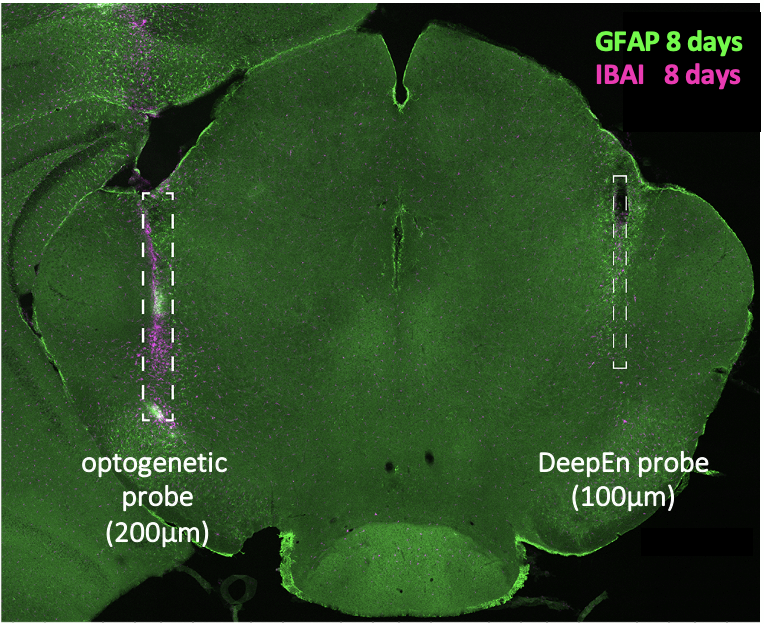
Minimal tissue damage
Insertion of our unique single-fibre probe has minimal effect on surrounding tissue and minimal effect on the physiological brain function.
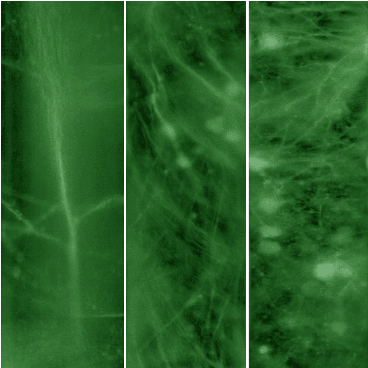
Subcellular resolution
Our technology enables single cell imaging and imaging of subcellular structures like axons, dendritic spines or buttons for studies of neuronal plasticity and connectivity.
Deep tissue penetration
Our 100 µm probe is stable enough to allow smooth insertion into deep and sensitive tissue regions that are currently not accessible with other technologies.
NeuroDeep® Microendoscopy Platform
Powerful bioimaging through a single fibre endoscope.
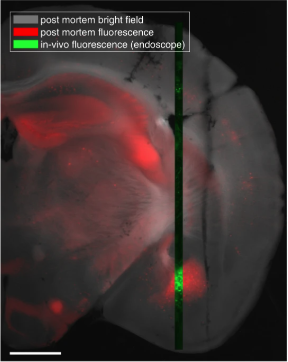
DeepEn’s unique NeuroDeep platform enables laser-scanning fluorescence imaging at the tip of a multimode fibre probe (⌀100μm) at any depth in living tissue with minimal structural damage and thus minimal effect on physiological function. Relying on advanced holographic technology, the device is capable of random-access observations across a 100 μm diameter field of view with submicrometric lateral resolution and on-the-fly adjustment of the focal distance. A read-out speed of up to 25,000 focal points per second allows for high-speed recording of neuronal activity. The instrument is a powerful tool for neuroscience, biomedical research and pharma laboratories.

Modalities
High-Resolution Imaging
Achieves submicrometre resolution of 0.7 µm, adhering to the Abbe limit for probes with 0.37 NA, operating at a wavelength of 491 nm.
Reduced Probe Footprint
The probe’s thickness of only 110 µm minimizes tissue damage, allowing for direct, image-guided insertion and immediate imaging.
Adjustable Focal Distance
Allows imaging across a set of preselected focal distances ranging from 0 to 50 µm from the probe facet.
Structured Illumination
Incorporates patterned illumination of user-selected areas for enhancing data acquisition rate to a kilohertz level.
Random-Access Scanning
25,000 focal points per second for swift selective scanning with no delays, regardless of the relative distance between scanning regions.
Excitation Wavelengths
Offers the flexibility of using either 491 nm or 532 nm excitation wavelengths, catering to a broad range of labelling options. Other wavelengths are possible on demand.
Multicolour Detection
Features simultaneous recording and visualization of different spectrally-separated fluorophores, excitable at the working wavelength.
Field of View
The field of view is spanning 100 µm in diameter and thus allows for imaging of multiple neurons across the fibre facet.
Applications
1) Structural Imaging
Imaging modality with full resolution at slow frame rate. The user can choose a slower scanning rate to achieve the submicron resolution to image individual cells, or small structures like dendrites and spines or even sub-cellular compartments.

2) Functional (Ca2+) imaging
Imaging with fast frame rate at low spacial resolution. Users can switch to this imaging modality for recording neuronal activity. The required higher frame rate is achieved by decreasing the spacial resolution, focussing only on the selected readout areas.


3) Blood flow velocity tracking
Measuring blood flow velocity in individual blood vessels for application in stroke research. Traces of individual red blood cells can be imaged (vasculature stained with FITC-dextran).


Source: Stibůrek, M., Ondráčková, P., Tučková, T. et al. 110 μm thin endo-microscope for deep-brain in vivo observations of neuronal connectivity, activity and blood flow dynamics. Nat Commun 14, 1897 (2023). https://doi.org/10.1038/s41467-023-36889-z

Discover with microendoscopy what has never been seen.
Real-time recording of active neuron structures during insertion of a NeuroDeep probe, visible on the user interface.
- Study in anaesthetised mouse model (Thy1-GFP line).
- Depth 5 mm – level of the amygdala
- Field of view: approx. 100×100 μm
Neuroscientific Background
In-vivo imaging through single Fibres
Holographic endoscope technology is an impactful innovation from the field of Neurophotonics. It is based on years of rigorous research conducted at renowned institutes, and was validated for deep-brain imaging in multiple independent laboratories.


Holographic Microendoscopy
This innovative technology turns a single multimode optical fibre into a laser-scanning microscope with much less traumatic application in-vivo and superior imaging performance.
The technology is based on precise control of light transport through optical fibres. During an initial calibration process, the complex light propagation through the fibre is characterised (transmission matrix „TM“ of the fibre) to achieve a focal point at the distal end of the fibre. The focal point scans over the sample, inducing emission of fluorescent light, which is collected back through the fibre and used to generate a microscopic image.
Publications
2012

2015

2018

2018

2023

News and Events
DeepEn GmbH Secures Seven-Figure Seed Round and Prestigious EIC-Transition Grant for Novel Holographic Endoscopes
DeepEn GmbH, a pioneer in holographic endoscopy...
DeepEn at FENS 2024: Bringing Holographic Endoscopes to the Heart of Neuroscience
Holographic Endoscope in Action! In June, the...
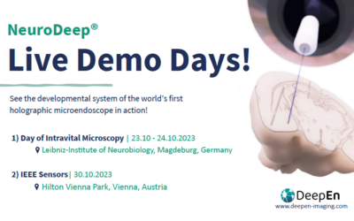
See NeuroDeep® in action at 2 Demo Days in October
Experience the developmental system of the...
Our Mission
DeepEn provides laboratories with powerful tools to study the deepest regions of the brain. Our mission is to support researchers in discovering, developing and applying the tools for prevention, diagnosis, and treatment of brain disorders.


DeepEn GmbH was funded as part of the EXIST-Transfer of Research program from 01.04.2024 to 30.11.2024.
DeepEn was founded in 2024 is a research transfer spinoff from Leibniz-Institute of Photonic Technologies
© 2025 DeepEn – All rights reserved
