
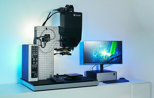
Introducing DeepEn One™ Microendoscopy Platform
Single-Fibre Endoscope for High-Resolution In Vivo Bioimaging
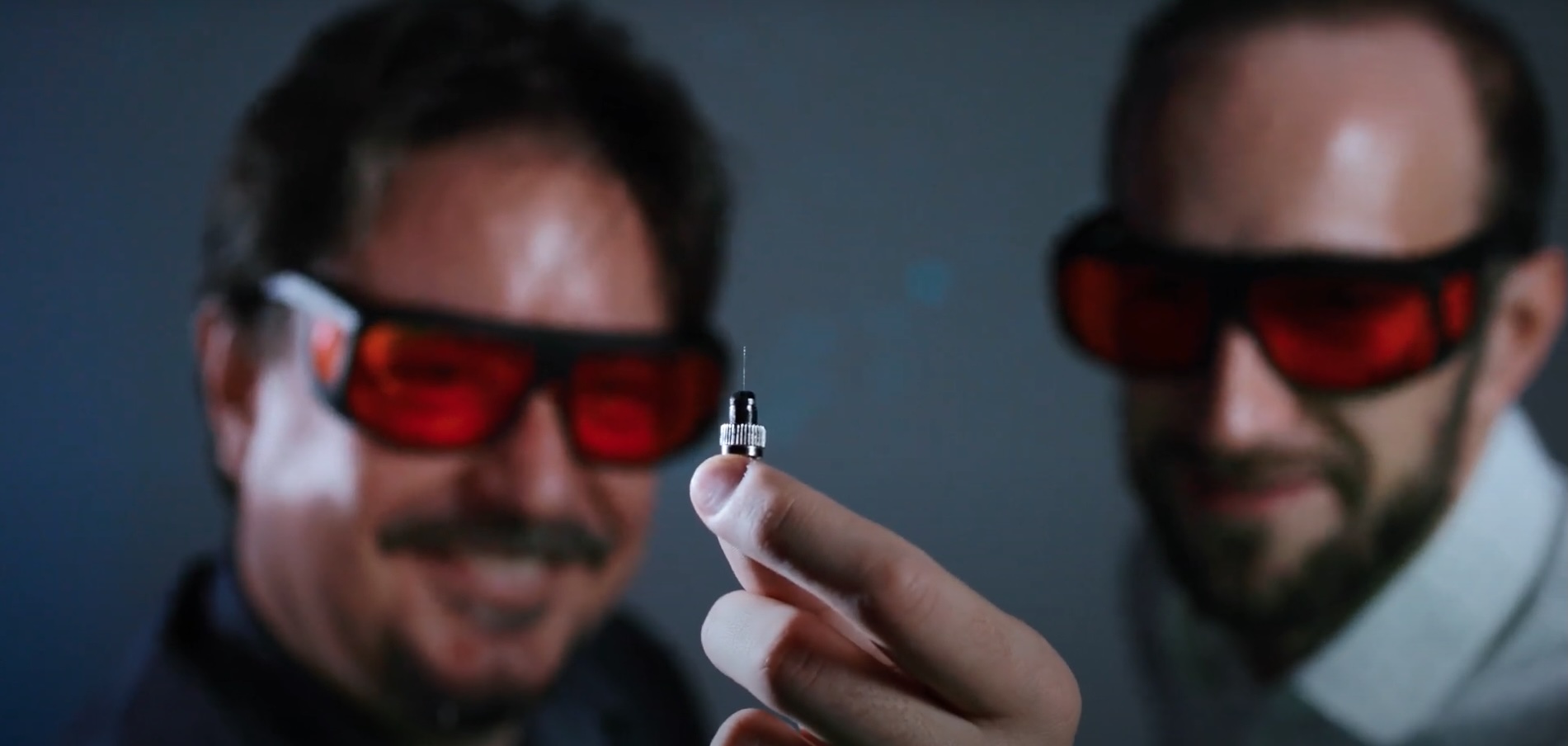
DeepEn One™ is a cutting-edge microendoscopy platform that delivers powerful in vivo bioimaging through a single multimode fibre endoscope (⌀100 µm). It enables researchers to probe deep into the living brain—reaching regions such as the amygdala, VTA, medial septum, brainstem, or spinal cord—while minimizing structural damage and preserving physiological function.
Discover the OneDiscover with microendoscopy what has never been seen.
Real-time recording of active neuron structures during insertion of a DeepEn One probe, visible on the user interface.
- Study in anaesthetised mouse model (Thy1-GFP line).
- Depth 5 mm – level of the amygdala
- Field of view: approx. 100 μm × 100 μm
From scientists
for scientists
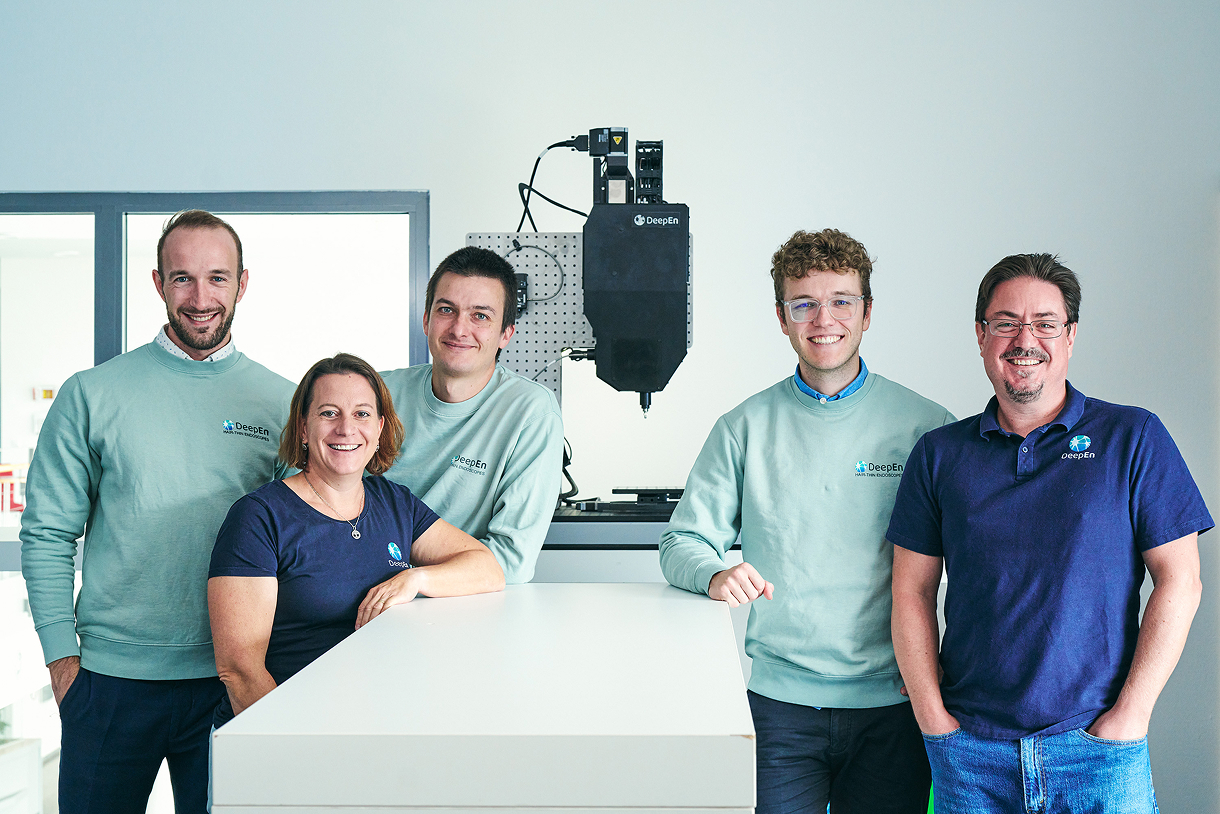
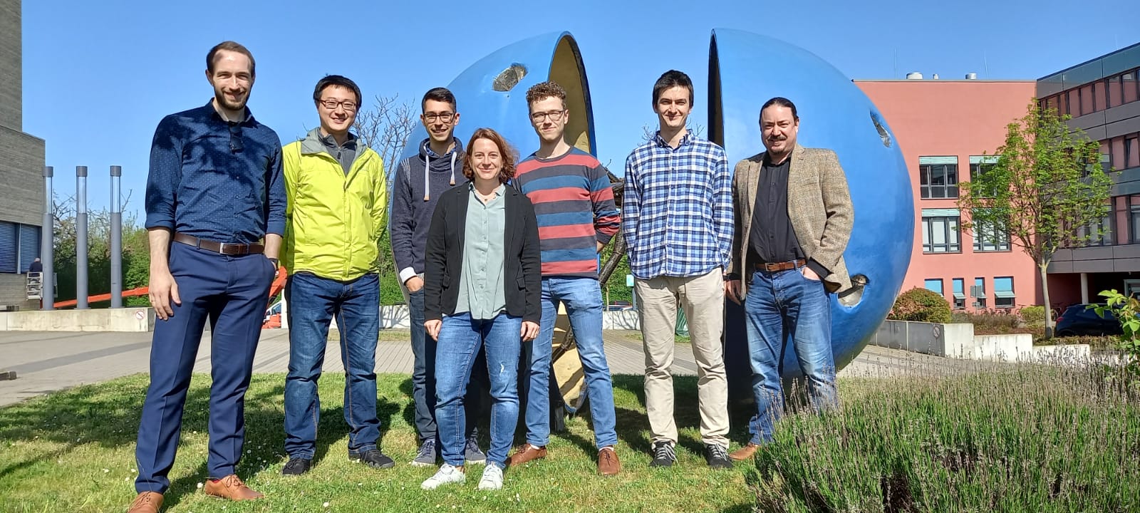
At DeepEn, we are a team of passionate scientists, engineers, and innovators united by a shared mission: to empower the scientific community with cutting-edge tools that make deep tissue imaging possible and more insightful. Born from years of hands-on research experience, our solutions are designed by scientists who understand the real challenges faced in the lab. We bridge the gap between complex technology and practical application — creating tools that let scientists focus on discovery, not the limitations of their instruments.
About DeepEnNews
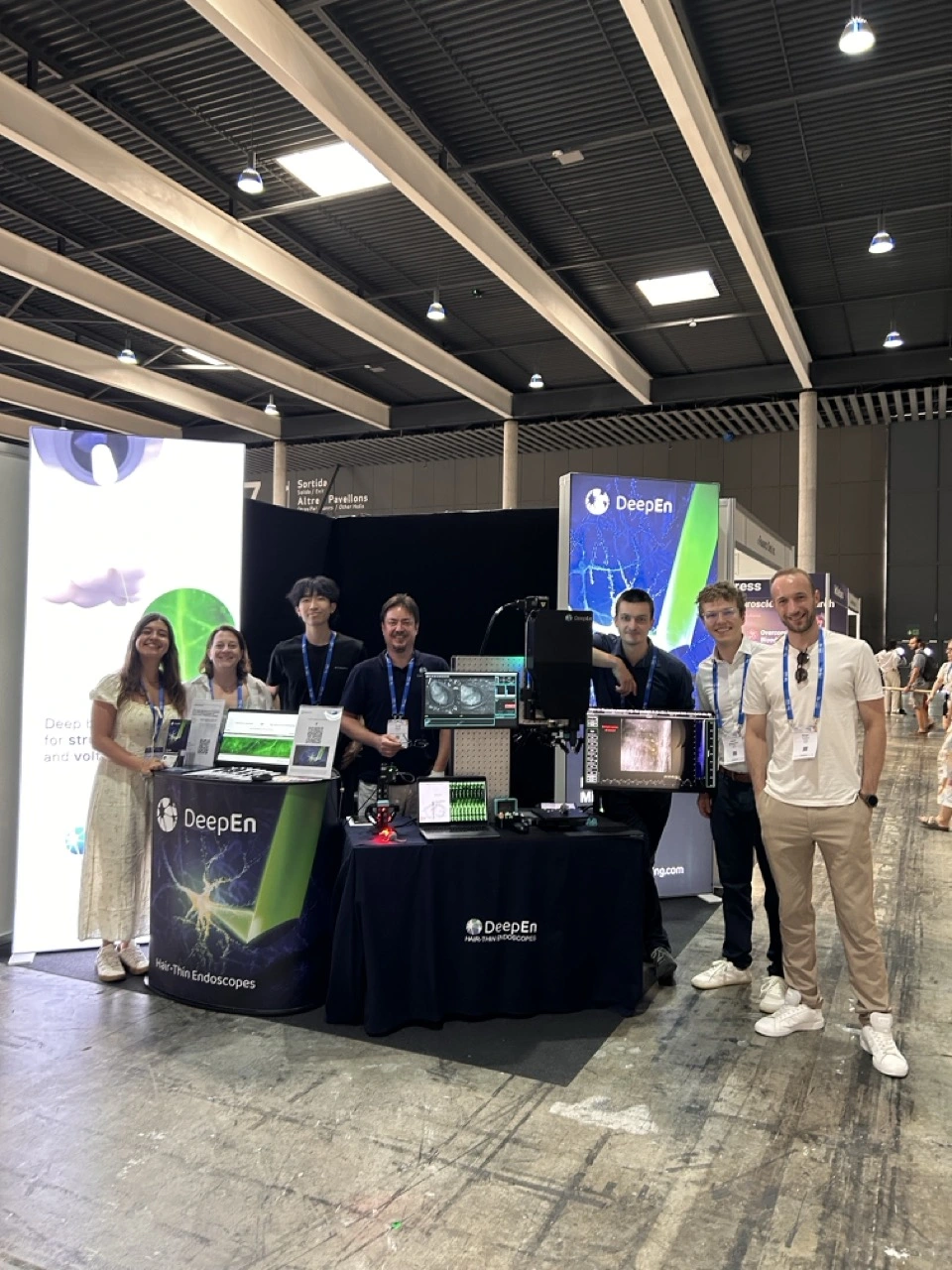
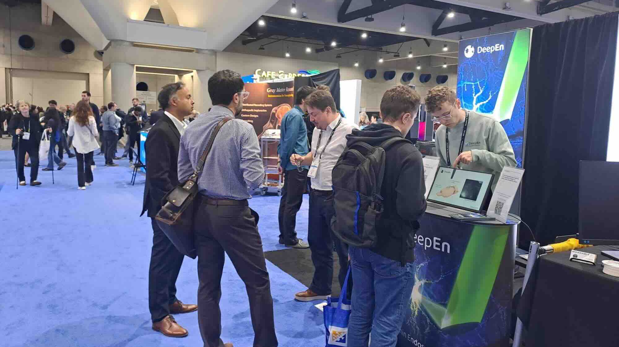


This Summer, DeepEn goes to
Barcelona for FENS 2026
We are excited to have a booth at FENS Forum 2026 in Barcelona, Spain from 6–10 July 2026. More details will be announced soon. Drop by, say hello, and discover our ultra-thin microendoscopes. Our team will share insights into the latest technological developments and research applications, and will be happy to discuss how the technology can support advanced in vivo neuroscience studies. We look forward to meeting you there!
More about FENS
Starting a new project? Or just curious to find out what you can do with an DeepEn One™ system?
Just email us and we will get back to you! Feel free to contact us also by phone.
Contact Hana