DeepEn One™
Application
Voltage imaging
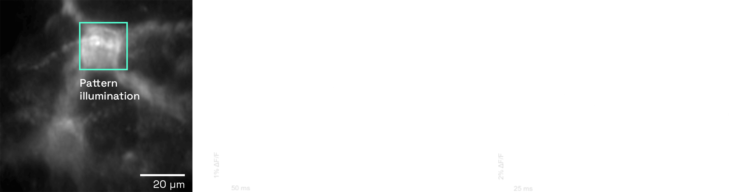
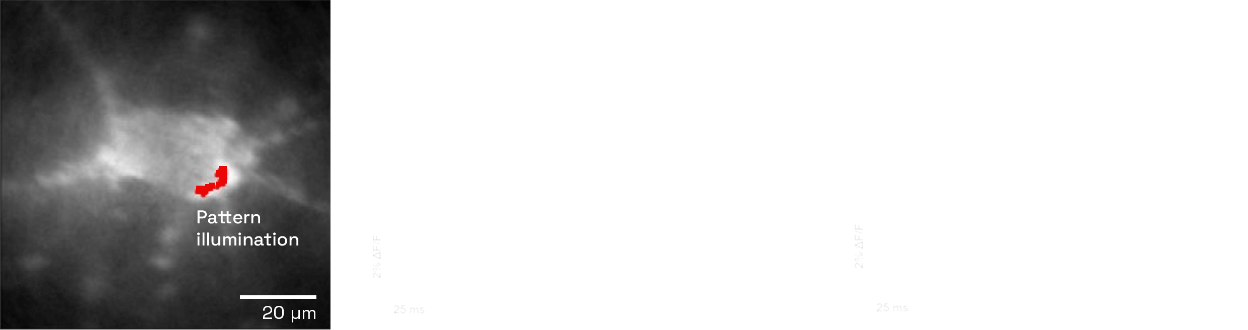
DeepEn One™ offers fast-frame-rate mode, suitable for voltage imaging, to record action potentials and subthreshold events for capturing neuronal activity in real time. By reducing spatial resolution and focusing on selected readout areas, this mode achieves the speed necessary to resolve spiking and subthreshold voltage activity.
It provides a tool for research into neuronal firing and cell signalling.
Cortex
Amygdala
Experiment Voltage recording with Voltron ST
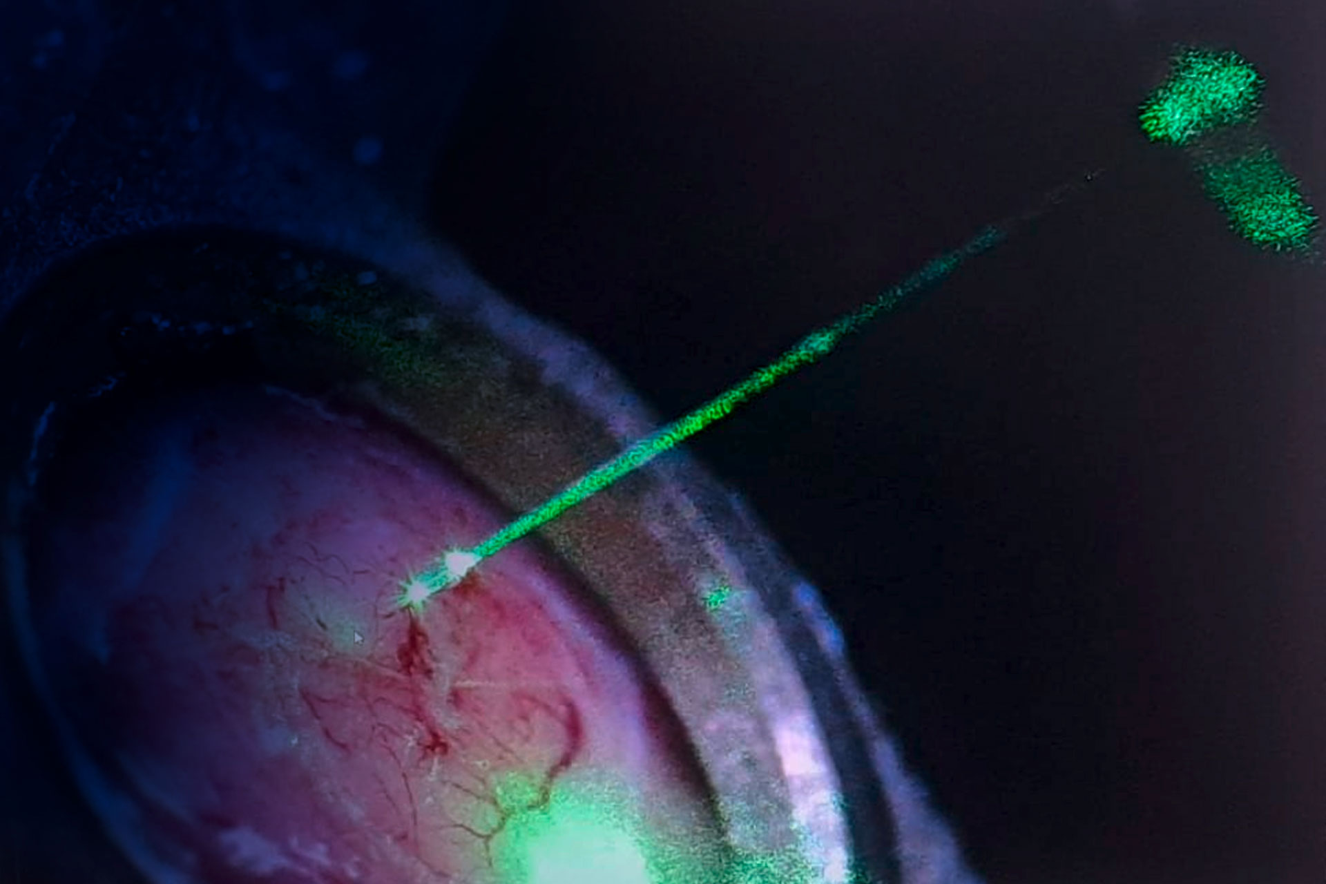
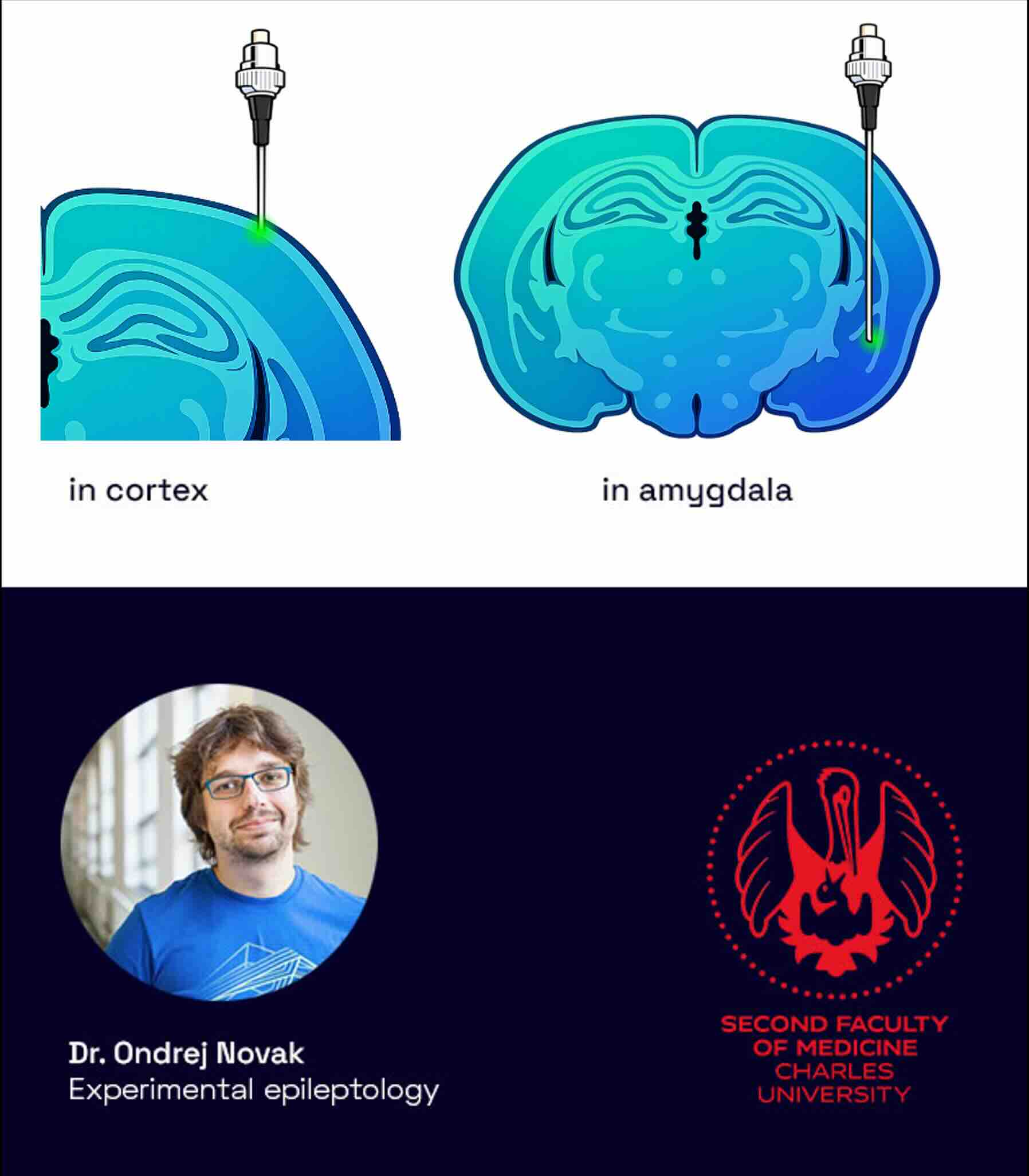
Voltage recordings were performed using the voltage sensor Voltron ST, labeled with JF525-HaloTag to enable fluorescence detection, at depths of 500 µm (cortex) and 5 mm (amygdala) in virally transfected animals. Voltage imaging in deep brain structures of mice models is challenging due to the combination of factors like the need for high speed (kilohertz) to capture rapid electrical signals, poor signal-to-noise ratios from tissue scattering and background fluorescence, and the difficulty of getting optical access to deep, hard-to-reach areas.
V1 cortical neurons and amygdala neurons were targeted and imaged under 532 nm excitation.
The experiments were conducted in collaboration with Dr. Ondřej Novák at the Experimental Epileptology Laboratory, Second Faculty of Medicine, Charles University.
- depth: 500 µm, resp. 5 mm
- animal: viral transfection
- voltage sensor: Voltron ST + JF525-HaloTag
- cells labelled and location: V1 neurons in cortex, amygdala
- excitation wavelength: 532 nm
Voltage imaging in ventral tegmental area
Dopaminergic neurons in VTA
Feasibility test –successful VTA voltage recording – subthreshold activity detectable

Experiment Overview
Voltage imaging was performed in the ventral tegmental area (VTA) to monitor dopaminergic neuron activity at depths of 4.5–4.8 mm. Dopaminergic neurons were labeled via viral transfection with novel genetically encoded voltage indicator ASAP6b* and imaged using 488 nm excitation. This feasibility test marks successful voltage recording in the deep, difficult to reach VTA, achieving detection of subthreshold neuronal activity .
Experiments were conducted under the supervision of Prof. Sebastian Haesler at the Novelty, Exploration & Curiosity Lab (NERF, Leuven, Belgium).
- depth: 4.5-4.8 mm
- animal: viral transfection
- fluorophore: Genetically encoded voltage indicator ASAP6b*
- cells labelled and location: dopaminergic neurons in VTA
- excitation wavelength: 488 nm
* GEVI ASAP6b was provided by Michael Lin’s laboratory, Stanford.
Improving positively tuned voltage indicators for brightness and kinetics
Sungmoo Lee, et al. bioRxiv 2024.06.21.599617; doi: https://doi.org/10.1101/2024.06.21.599617

Starting a new project? Or just curious to find out what you can do with an DeepEn One™ system?
Feasibility test –successful VTA voltage recording – subthreshold activity detectableJust email us and we will get back to you! Feel free to contact us also by phone.sdfsdf
Contact Hana