DeepEn One™
Application
Structural imaging
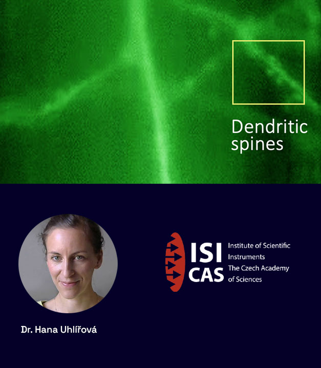
DeepEn One™ enables high-resolution structural imaging . Users can achieve submicron resolution- ideal for visualizing individual cells, and even fine neuronal structures such as dendrites and spines. This high-resolution imaging capability does not degrade with depth. This mode provides detailed structural information essential for studying cellular morphology and connectivity at the microscale.
Imaging the whole depth of mouse brain from cortex to amygdala neurons and processes with subcellular resolution
Video of the whole depth of mouse brain from cortex to amygdala neurons
Experiment Overview
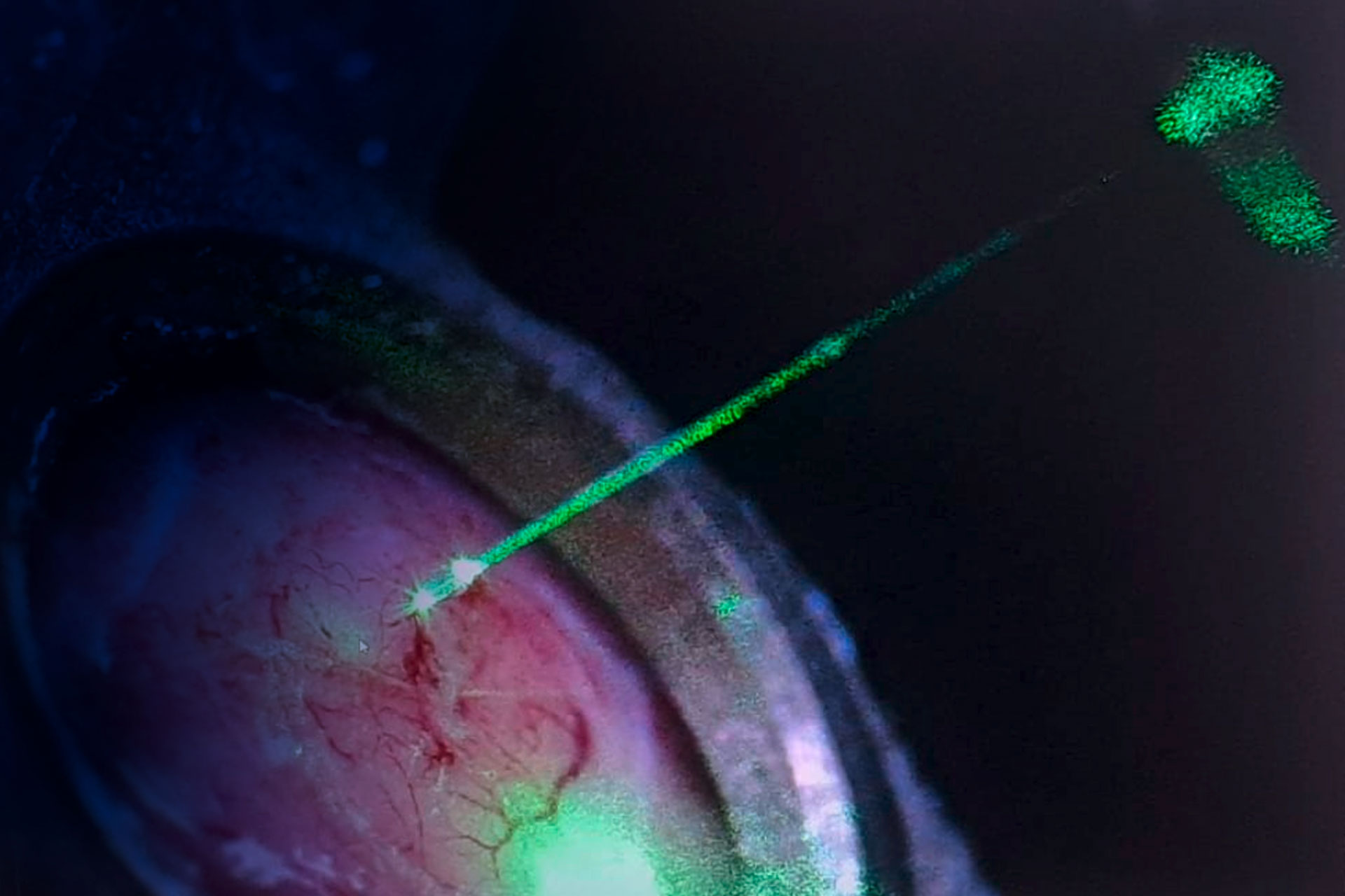
Whole-depth imaging of the mouse brain (0–5 mm, from cortex to amygdala) was performed in transgenic Thy1-GFP mice expressing GFP in subsets of neurons and their processes. Using 488 nm excitation, neuronal somata, dendrites, axons, and subcellular structures were visualized with high spatial resolution along a single insertion path (live imaging during insertion, images then post-processed into one block).
This approach enabled continuous mapping of neuronal morphology and connectivity from the cortical surface through hippocampal layers down to the amygdala within the intact mouse brain.
Imaging was conducted with at the Complex Photonics Laboratory of the Institute of Scientific Instruments (ISI), Czech Academy of Sciences, Brno.
- depth: 0 – 5mm
- animal: transgenic Thy 1 mice
- fluorophore: GFP
- cells labelled and location: from cortex to amygdala
- excitation wavelength: 488 nm
Imaging in ventral tegmental area
Dopaminergic neurons with subcellular resolution
Experiment Overview
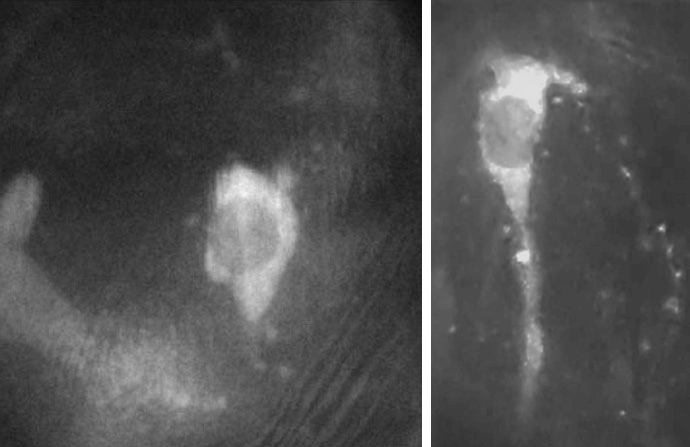
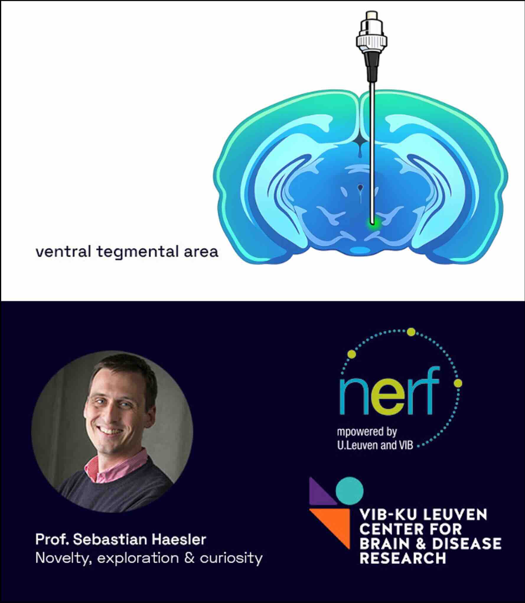
Imaging was performed in the ventral tegmental area (VTA) to visualize dopaminergic neurons with subcellular resolution at a depth of 4.5–4.8 mm. Neurons were labelled via viral transfection to express the calcium indicator GCaMP8s – for the purpose of calcium imaging, in dopaminergic cells within the VTA and imaged using 488 nm excitation laser.
This feasibility test represents the first successful imaging of the VTA, achieving subcellular resolution with detectable neuronal activity in a single animal.
The experiments were conducted in collaboration with Prof. Sebastian Haesler at NERF lab (Neuroelectronic research Flanders, Leuven, Belgium).
- depth: 4.5 – 4.8 mm
- animal: viral transfection
- fluorophore: GCaMP8s for calcium imaging
- cells labelled and location: dopaminergic neurons in VTA
- excitation wavelength: 488 nm

Starting a new project? Or just curious to find out what you can do with an DeepEn One™ system?
Just email us and we will get back to you! Feel free to contact us also by phone.
Contact Hana