DeepEn One™
Application
Calcium Imaging
DeepEn One™ offers fast-frame-rate mode suitable for calcium imaging for capturing neuronal activity in real time. By reducing spatial resolution, this mode achieves the speed necessary to monitor dynamic calcium signals in labelled neurons.
It provides an efficient solution for studying neuronal signalling, network dynamics, and functional connectivity deep in living tissue.
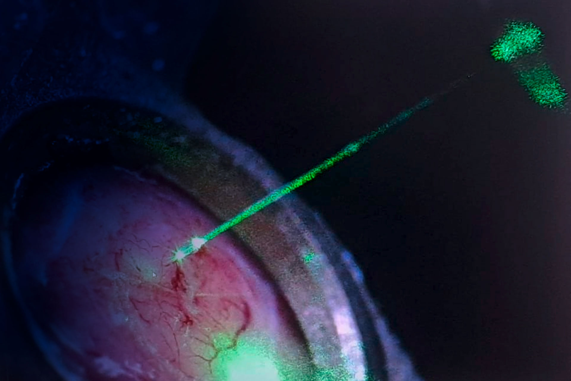
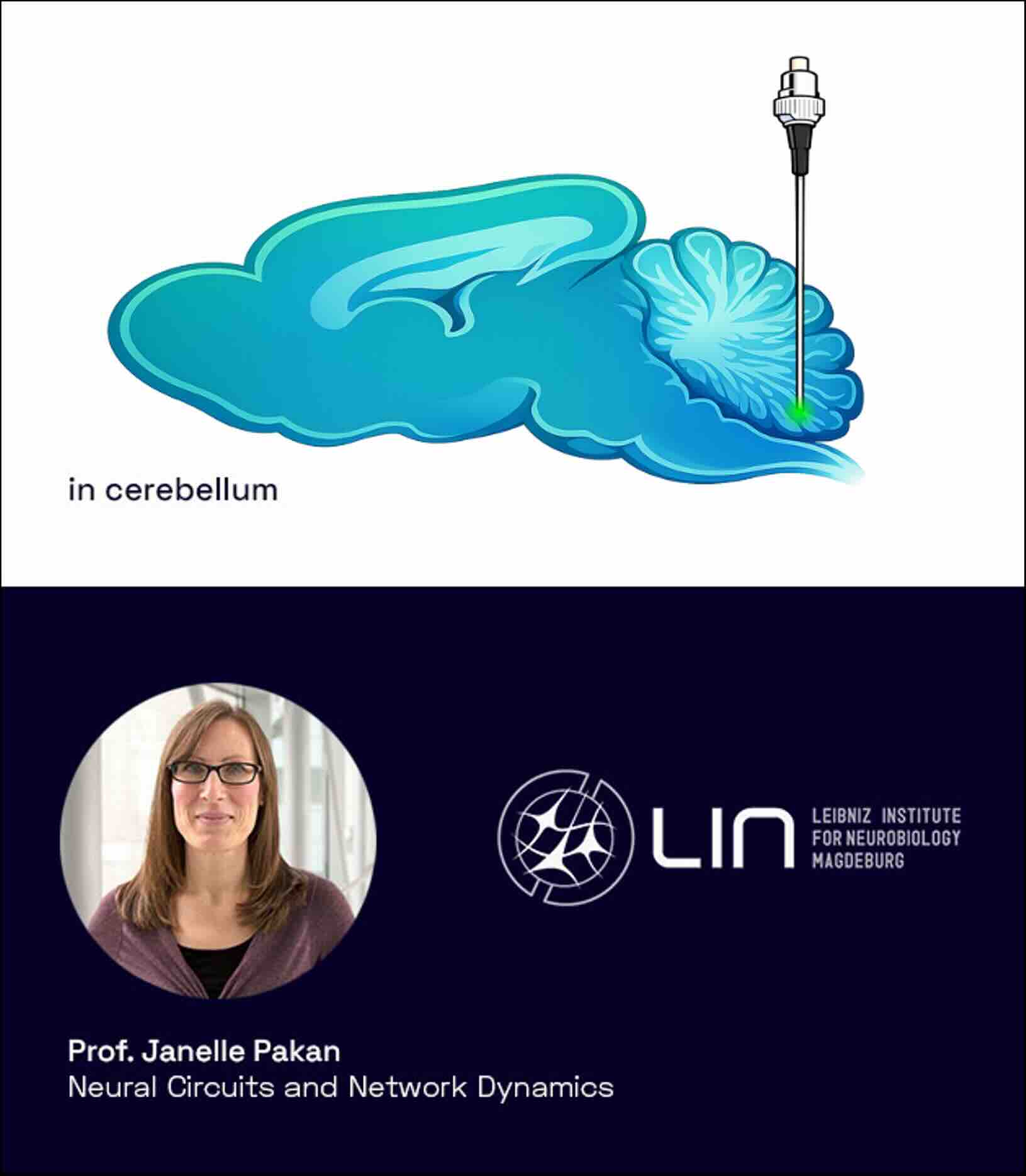
Calcium imaging in cerebellum
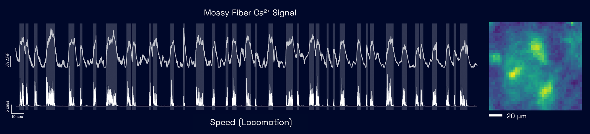
Recording of calcium signals of mossy fibres deep in cerebellum during locomotion
Correlation between movement and calcium signals
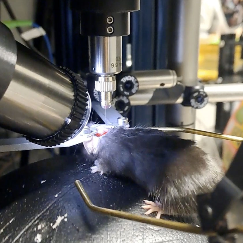
Experiment Overview
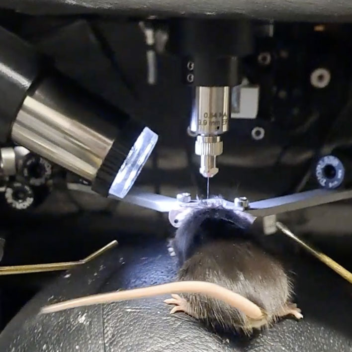
Calcium imaging was conducted in Thy1-GCaMP6m mice to record mossy fibre activity deep within the cerebellum (>3 mm) during locomotion. Investigating the cerebellum in mice is technically challenging due to its small size, layered organization, and location at the back of the brain, between the ears—making it inaccessible to conventional two-photon microscopy. Using 488 nm excitation, calcium signals were recorded from mossy fibres in the cerebellar grey matter through a miniaturized endoscopic setup. Mice were imaged while walking on a PhenoSys JetBall system to correlate mossy fibre activity with movement.
Prof. Janelle Pakan (Neural Circuits and Network Dynamics Lab at Leibniz Institute for Neurobiology) is interested in how cerebellum is involved in processing sensory and motor inputs into behavioural outputs, and how this changes with learning.
- depth: 3+mm
- animal: Thy1 mice
- fluorophore: GCaMP 6 M
- cells labelled and location: mossy fibres in cerebellum
- excitation wavelength: 488 nm
Mice were imaged while walking on a PhenoSys JetBall system to correlate mossy fibre activity with movement.
When working with animals, DeepEn and its partners follow the highest animal welfare standards under the 3R framework (Directive 2010/63/EU on the protection of animals used for scientific purposes).

Starting a new project? Or just curious to find out what you can do with an DeepEn One™ system?
Just email us and we will get back to you! Feel free to contact us also by phone.
Contact Hana