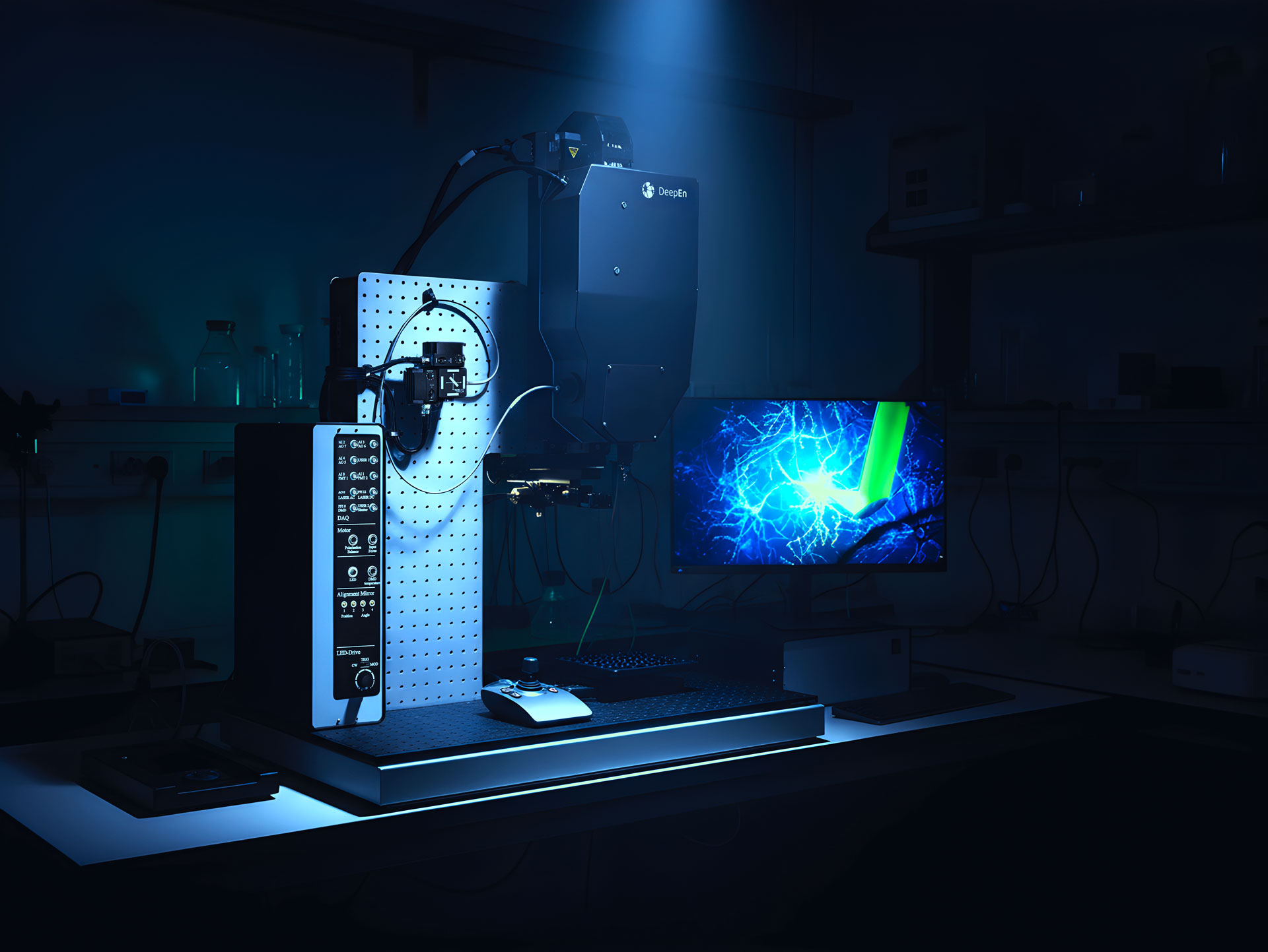
DeepEn One™ in vivo
Microendoscopy Platform
Powerful bioimaging through a single fibre endoscope.
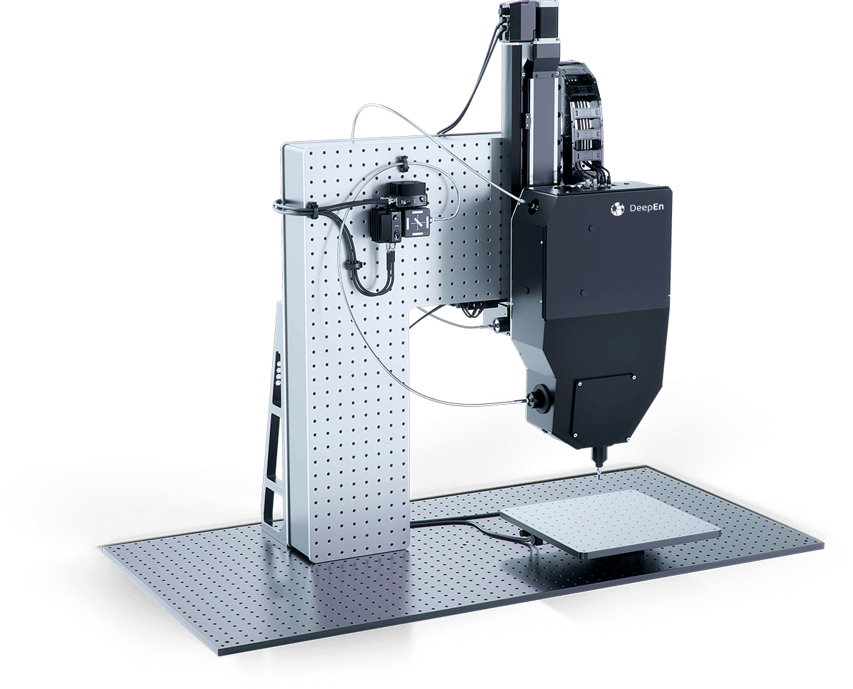
DeepEn One™
Microendoscopy Platform
Single-Fibre Endoscope for High-Resolution in vivo Bioimaging
DeepEn One™ is a cutting-edge microendoscopy platform that delivers powerful in vivo bioimaging through a single multimode fibre endoscope (⌀100 μm). Combining laser-scanning fluorescence microscopy with holographic wavefront control, it enables researchers to probe deep into the living brain—reaching regions such as the amygdala, VTA, medial septum, brainstem, or spinal cord—while minimizing structural damage and preserving physiological function.
With submicron lateral resolution and a 110 µm diameter field of view, DeepEn One™ provides random-access imaging and real-time focus adjustment. Its ultra-fast acquisition rate of up to 25,000 focal points per second makes it ideally suited for fast neuronal activity recording, including studies based on voltage imaging.
Workflow with DeepEn One™
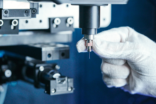
Probe mounting
Attach the hair-thin probe to the DeepEn One core. A self-aligning fibre connector ensures easy mounting and precise, repeatable positioning of the probe tip.
Calibration
Before each imaging session, DeepEn One™ automatically aligns the fibre tip with the calibration module.
The software guides the user through the process, which takes approximately 10 minutes.
Navigation
Using a joystick or GUI, the fibre is guided to the craniotomy and inserted into the target region.
Additional navigation module can be used for high-resolution visualisation, enabling precise positioning and alignment with brain-atlas references.
Imaging
After calibration, the focal point scans across the selected plane near the fibre tip, exciting fluorescence, which is collected through the same fibre.
Scanning parameters – sampling density, focal distance, and random-access scanning, are adjustable via the GUI.
Key Areas of Research
- Fundamental neuroscience: neuronal plasticity, connectivity mapping, calcium and voltage imaging, microcircuit analysis
- Disease research: Alzheimer’s, Parkinson’s, psychiatric disorders (depression, anxiety, schizophrenia), addiction, chronic pain, epilepsy, sleep and arousal studies.
- Biomedical discovery & preclinical research: drug testing, mechanism-of-action analysis, and biomarker validation.
Why DeepEn One™?
This innovative single-fibre endoscope provides laboratories with a compact, high-performance platform for exploring brain function and cellular processes at unprecedented depth and resolution—bridging fundamental discovery and translational neuroscience.
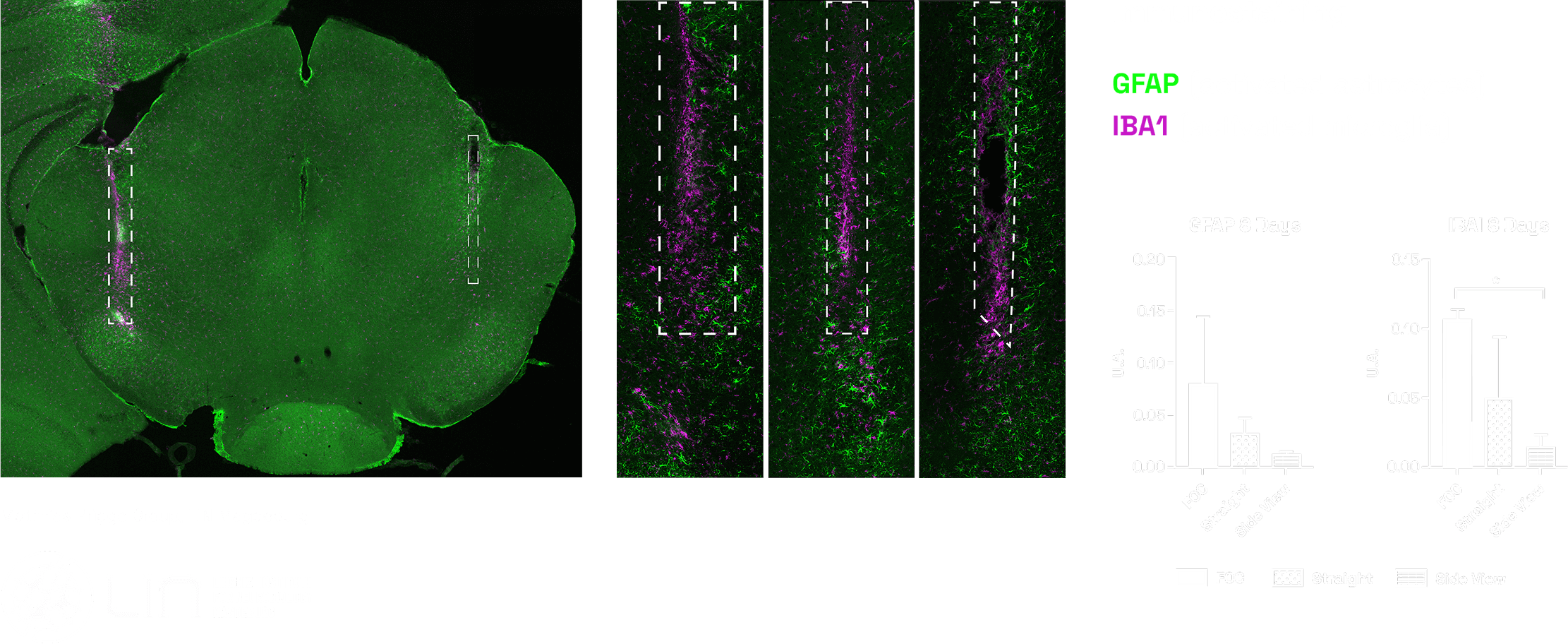
Minimal Invasiveness & Deep Penetration
The stability of our 100-µm-probe allows for smooth insertion into deep brain regions with minimal effect on the tissue or the function.
Postmortem tissue analysis shows that the 110-µm-DeepEn-probe causes significantly less insertion-induced inflammation and tissue damage. Even compared to a standard 200 µm optogenetic or photometry probe, the DeepEn fibre exhibits minimal impact on brain tissue.
DeepEn One™ enables high-resolution imaging at any depth within the brain, reaching even the deepest and most sensitive neural structures.
High spatial and temporal resolution
By using holographic control of light in multimode fibres, DeepEn One™ enables imagng at any depth across the brain, with the ability to switch between high spatial and high temporal resolution modes during a single session.
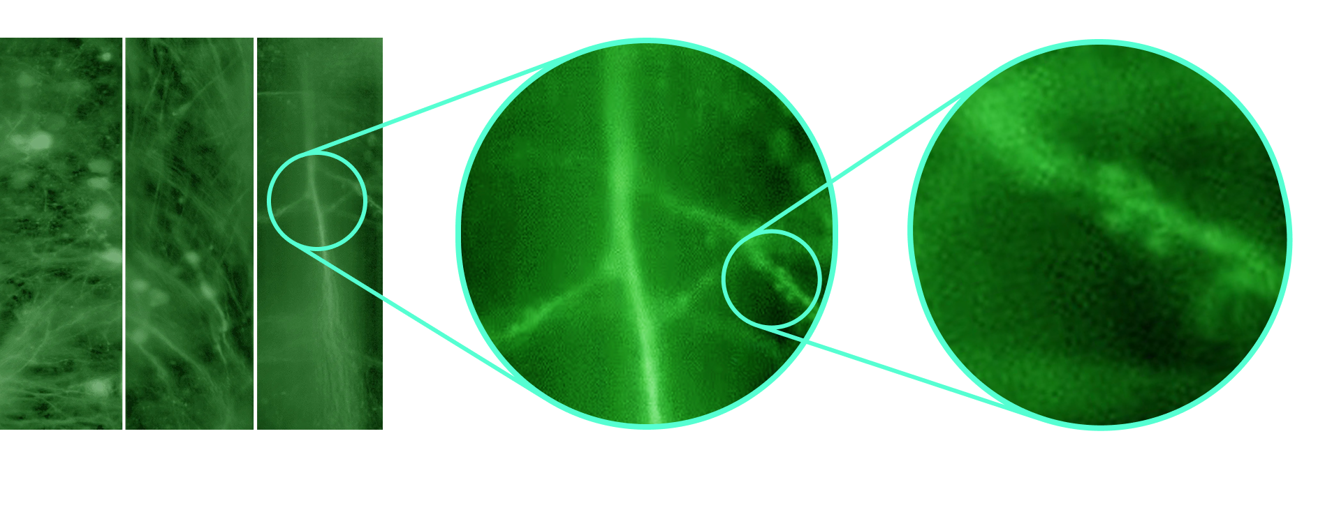
High Spatial Resolution – Seeing the Brain in Unprecedented Detail
DeepEn One™ delivers exceptional spatial resolution, enabling precise imaging of neuronal structures deep within living brain tissue. By combining advanced holographic wavefront shaping with hair-thin optical probes, it resolves fine subcellular features at any depth across the brain.
This allows researchers to visualize complex neuronal networks and synaptic dynamics, unlocking new possibilities for in-vivo neuroscience.
High Temporal Resolution – Recording neuronal activity
DeepEn One™ delivers high temporal resolution imaging, capturing rapid neuronal dynamics in real time. Leveraging fast holographic modulation, the system enables millisecond-scale monitoring of neuronal activity deep within scattering tissue.
This performance allows researchers to record Calcium and even Voltage signals, providing unprecedented insight into the neuronal signalling, network dynamics, and functional connectivity deep in living brain.
Modalities

Multicolour operation
Features simultaneous recording and visualization of spectrally-separated fluorophores, excitable at the working wavelengths.

Adjustable Focal distance
Provides dynamic refocusing over a 0–50 µm depth range from the probe facet, enabling volumetric imaging.
Random access scanning
Scans up to 24,000 focal points per second, enabling rapid, user-defined sampling anywhere in the field of view without motion delays.

Structured illumination
Incorporates patterned illumination of user-defined regions to enhance data acquisition rates up to the kilohertz range.
Technical specifications:
| Specification | Description |
|---|---|
| Fibre size / field of view (FOV) | ∅ 110 µm / ∅ 100 µm |
| Excitation wavelength | Blue 488 nm or Green 532 nm (450, 471, 488, 515, 532, 561 nm) two-colour excitation possible |
| Submicron optical resolution | Equivalent to 20× microscope objective |
| Focal plane adjustment | Up to 50 µm from the fibre facet |
| Scanning rate | 24,000 scanned points per second – Full FOV: 0.24 fps – 1000 points: 24 Hz – 24 points: 1 kHz |
| Pixel acquisition time adjustable (pixel dwell time) | 40 µs – 10 ms, pixel-based averaging |

Would you like to get to know us, invest in DeepEN or become part of our visionary team?
Just email us and we will get back to you! Feel free to contact us also by phone or write us a message via the contact form.
Contact Hana